Article
Scanner
3m read
Tips For Efficient Scanning In Full Arch Cases
Achieve fast, accurate full arch scans with optimal scanner positioning, and streamlined protocols.
Dr. Amr Adel
on
Jul 31, 2024
Tips For Efficient Scanning In Full Arch Cases
In a fully digital workflow, using an intraoral scanner (IOS) streamlines the entire process from start to finish. The data captured directly from the patient’s mouth is sent to computer-aided design (CAD) software for the creation of prosthetic restorations, which are then transferred directly to a milling machine for manufacturing. This digital approach eliminates common problems associated with traditional methods, such as material distortion, patient discomfort due to gag reflex or allergies, and the risk of infection transmission between the patient and technician.
Intraoral scanners provide numerous advantages, including increased patient comfort, faster scanning times, and reduced storage and transportation needs. When working with implants and prostheses, achieving a completely digital workflow requires the use of intraoral scan bodies (ISBs). However, several factors can impact the scanner's performance, such as saliva, movements of the tongue and cheeks, the characteristics of the edentulous ridge, and the position and angulation of implants. Ensuring a passive fit between the prosthetic framework and implants is essential to avoid mechanical and biological complications, which are crucial for the long-term success of restorations.
Full arch scanning can be a challenging process, particularly in fully edentulous cases where accuracy and efficiency are key. Here are some essential tips to make your full arch scanning process more efficient, especially when using the Atomica S200 intraoral scanner.

Preparing the Patient
Before beginning any scan, it’s essential to prepare the patient properly. Proper patient positioning is crucial for achieving accurate and efficient full arch scans, particularly in challenging cases like edentulous or partially edentulous arches. Begin by ensuring that the patient is comfortably seated in a position that allows the clinician unobstructed access to the entire oral cavity. The patient’s head should be slightly tilted back, with the chin slightly elevated, to create a direct line of sight and facilitate easy access to both upper and lower arches. Utilizing a comfortable headrest can help maintain this position throughout the scanning process, reducing the likelihood of unwanted movements that can compromise scan quality. Instruct the patient to keep their lips relaxed and mouth open gently to avoid excessive strain or discomfort.
Ensuring that the patient is calm and relaxed is also important; explain the procedure in detail beforehand, including how long it will take and what they can expect. A well-informed patient is less likely to move unexpectedly, allowing for a smoother and more efficient scanning experience. Overall, careful attention to patient setting, positioning, and comfort helps achieve a precise and comprehensive full arch scan, reducing the need for rescans and enhancing both clinical workflow and patient satisfaction.
Start with a Clear Field of View
To further enhance visibility and access, consider using cheek retractors to move soft tissues like cheeks and lips away from the scanning area, thereby minimizing the risk of soft tissue interference during the scan.
Controlling moisture in the oral environment is also critical—excessive saliva can cause reflections that distort scan data. Use a saliva ejector or gauze to keep the field dry and clear. Proper lighting in the operatory is another key aspect; ensure the room is well-lit, but avoid direct overhead lights that could create glare or shadows in the patient’s mouth.
Also, Effective lighting is a key factor in capturing high-quality scans. Ensure that the operatory is well-lit but avoid direct light that could shine into the patient’s mouth.

Maintaining a Systematic Scanning Path
Various scanning strategies have been developed to optimize the accuracy and efficiency of full arch intraoral scanning. The most common approach is the continuous scanning technique, where the operator moves the scanner in a fluid, uninterrupted motion across the dental arch. This method typically begins with the occlusal surfaces, followed by buccal and lingual aspects, often in a zigzag pattern or U-shape pattern. An alternative strategy is the segmental scanning technique, which divides the arch into discrete sections (e.g., quadrants or sextants) that are scanned individually and then stitched together by the software. This method can be particularly useful for challenging cases or when working with scanners that have a smaller field of view. The patch scanning technique involves capturing multiple overlapping images of small areas, which can be beneficial for capturing highly detailed regions or complex dental anatomy. Some clinicians employ a hybrid approach, combining elements of continuous and segmental scanning to adapt to specific clinical situations. Advanced strategies may incorporate specific patterns, such as the "steering wheel" technique for anterior teeth or the "wave" technique for posterior regions. Ultimately, the choice of scanning strategy depends on factors such as scanner technology, software capabilities, clinical scenario, and operator preference. Research has shown that while different strategies can yield comparable results in terms of accuracy, they may vary in scanning time and ease of use for the clinician.
Ready to elevate your scanning? Book a demo today!
Scanning Protocol for edentulous cases
Scanning a fully edentulous arch presents unique challenges due to the absence of distinct anatomical landmarks, which are essential for orientation and alignment in digital impressions. However, with the right protocol, precise and accurate digital impressions can still be achieved, providing a reliable foundation for fabricating implant-supported prostheses or complete dentures. Here are some tips for efficient edentelous scanning:
Tips for reliable Maxillary scans:
Start From a fixed anatomical landmark (Tuberosity or Ruggae area)
Make your way across the ridge with a steady and consistent pace
When scanning the palate, do so in a zigzag pattern to ensure smooth transition between the tissue.
Tips for reliable Mandibular scans:
Start From a fixed anatomical landmark (Ideally the retromolar pad)
Have a slight lingual inclination and move steadily towards the midline
Return to the retromolar pad with a buccal inclination
Pause the scanner, allow the patient to rest and remove the saliva with suction
Start from the midline and now work your way to the retromolar pad of the opposite side with a slight lingual inclination
Now finish returning to the midline with a slight buccal inclination at a steady rate
Note that sometimes resorbed lower ridges have extremely mobile tissue, which leads to a double image if the same area is scanned twice with a different position in each time
To avoid this, the tissue must be immobilized either by:
The clinician while scanning manually
Special retractors that retract the tissue while fixating them


Scanning strategy not only saves time and effort, more and more research is coming out to support that certain strategies offer improved accuracy, especially in edentulous jaws.
Jamjoom et al in 2024 compared 6 different scanning strategies and their effect on accuracy, They concluded that the strategy did in fact have an effect an accuracy, the most accurate being P-O-B: Starting posteriorly and proceeding along the palatal or lingual aspect of the ridge, returning along the occlusal aspect, and finally scanning the buccal aspect(Figure 3-B).
This supports the strategies discussed earlier, that indicate starting with a fixed anatomical reference point is key to a proper edentulous scan.

Ready to elevate your scanning? Book a demo today!
Techniques to increase the accuracy of edentulous arch scanning
Artificial Landmarks to Improve the Accuracy of IOSs
Artificial landmarks are strategically placed markers used to enhance the accuracy of intraoral scanners (IOSs) when capturing edentulous arches. These markers can be radiopaque or adhesive fiducials, small dots, or lines placed on the edentulous ridge, dentures, or scanning stents. They serve as reference points, aiding the scanner in maintaining orientation and minimizing errors during image acquisition and stitching. By providing consistent, identifiable features, artificial landmarks help reduce distortions that commonly occur in the soft tissue scanning of edentulous arches. They also facilitate the alignment of multiple scans, particularly when merging intraoral scans with CBCT data, resulting in more precise digital impressions. The use of artificial landmarks is especially beneficial in cases with uniform soft tissue contours, where the absence of natural anatomical features can make accurate scanning challenging.
Papaspyradikos et al 2022, concluded that using self adhesive radiopaque fiducial artificial landmarks (Figure 4) in the absence of anatomical landmarks improved both the scanning process as well as the accuracy and fitting of the final restoration.

Splinting ISBs to Improve the Accuracy of IOSs
Splinting implant-supported bite splints (ISBs) is an effective technique to enhance the accuracy of intraoral scanners (IOSs) when capturing edentulous arches. This method involves stabilizing ISBs with rigid materials or frameworks to ensure they remain securely in place during the scanning process. By minimizing movement and ensuring a stable reference point, splinting helps to reduce distortion and improve the consistency of digital impressions. The splinting process involves fabricating a splint that fits over the edentulous arch and provides additional support to prevent displacement. This ensures that the IOS can capture accurate, high-quality data without the complications that can arise from the natural movement of the arch or prosthesis. Additionally, splinting ISBs can facilitate the precise alignment of intraoral scans with other imaging modalities, such as CBCT, by providing fixed reference points. This technique is particularly useful in complex cases where maintaining the exact position of the ISB is critical for achieving optimal scanning results.
It’s preferable that the splint be at least 4 mm away from the ridge, to not interfere with the ridge scan
The splint should be a different color than the gums
The splint should have a rough/dull surface that is slightly irregular so that it’s easily scanned.
Cappare et al in 2019 conducted a randomized clinical trial where conventional vs digital full arch rehabilitation techniques were compared to each other. They concluded that using splinted scan bodies and digital scans the accuracy is comparable to the conventional method, and has an advantage in terms of patient comfort and time

Maximizing the Use of Integrated Software Features
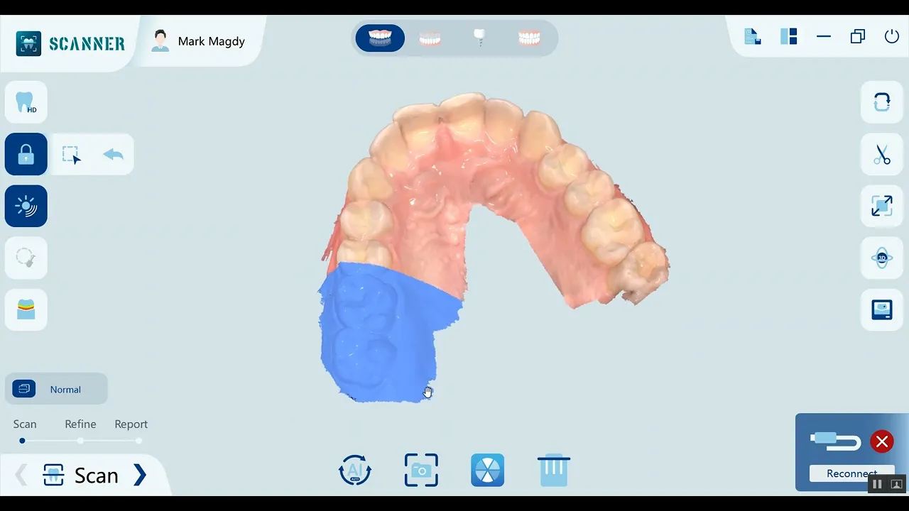
Atomica’s software has various features to aid in full arch cases, the fact that it is powered by AI enhances the ability to scan edentulous areas and metal surfaces significantly.
Here are some of the features that enable a seamless workflow are:
Lock Area:
Workflow: You can use this feature to lock a perfectly scanned area, so that no matter how long you rescan, the locked area remains unaffected.
Clinical application: locking a perfectly retracted and isolated subgingival margin line, so that if the gum collapses during preparation modifications and rescans, the scanned margin remains the same without the need for repeating retraction and isolation
Shining filter:
Workflow: The shining feature allows the scanning of metal objects to be significantly easier, reducing your implant workflow time drastically and ensuring all the minute details are scanned accurately to guarantee perfectly fitting final restorations.
Clinical applications: During full arch scans, if an implant/abutment is not being scanned completely, the shining filter is used to capture all the missing minute details.
Undercut checker:
Workflow: No need to rely on faulty visual inspection of the eyes anymore, using our undercut check tool, you simply adjust your scan view to be occlusal on the scan screen, and the software will automatically highlight the undercuts in a color code depending on their intensity, so you can just adjust them and rescan accordingly.
Clinical application: Used to check for parallelism between preparations and abutments.
Incorporating these tips into your full arch scanning routine can significantly enhance your efficiency and accuracy. By following proper scanning protocols, using the right equipment, and leveraging advanced features like AI-powered filtration and occlusion analysis, you can achieve optimal results even in complex cases. Start implementing these strategies today to streamline your workflow and elevate the quality of your digital impressions.
Ready to elevate your scanning? Book a demo today!
—————————————————————————————————


